

TMT-1 Arthritis – Case Study
Dr Lintz is a consultant orthopedic surgeon in France and an author on more than 70 scientific publications. He is a strong proponent of WBCT and 3D imaging within the foot and ankle community. Dr Lintz has been working with Disior® for over 18 months (at time of writing) and he regularly uses the analysis provided by our medical imaging software, Bonelogic®, to help with his patients. In this short case study, he explains how the 3D models and distance mapping analysis helped with diagnosing a subtle case of TMT1 arthritis.
Arthritis of the first tarsometatarsal (TMT-1) causes pain. It is commonly caused by trauma or primary instability, and affects many people that consult with foot specialists.
This patient presented with medial midfoot pain that had been ongoing for several months. I had a bilateral WBCT conducted to get an accurate 3D images of the patients’ anatomy under natural loading conditions.
The medical images were run through the Foot & Ankle module of Disior®’s Bonelogic® software and additional 3D analysis were performed by Eero Huotilainen, PhD.


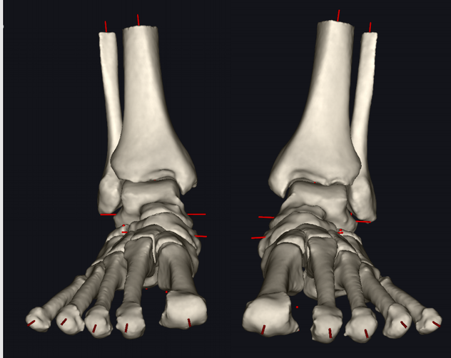
The accurate 3D models, revealed a very subtle but asymmetrical TMT1 arthritis on the left foot, where the metatarsal had slipped on the cuneiform (Figure 1).
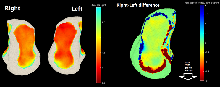
Figure 2. Right-left comparison of TMT1 joint space.
By comparing the joint spacing between the left and right TMT1 you can see the subtle narrowing of the left TMT1 joint indicative of arthritis (Figure 2).
Figure 3. Video of the distance mapping of the Lisfranc area.
Distance mapping of the Lisfranc region (Figure 3) showed subluxation and approximation in the dorsal aspect of the joint explaining the patient’s pain.
These insights allowed Dr Lintz to choose the most appropriate treatment, firstly a pair of supporting insoles, then a TMT1 fusion.
Thank you for taking the time to read this case study.
Disior® Bonelogic® was built specifically to help clinicians make diagnoses and plan patient treatment. Bonelogic® takes DICOMs from CT, CBCT or WBCT and generates accurate models and analytics that describe the relationships between bones, like those used in this case study.
If you’re interested in learning how Disior®‘s products can aid in your clinical practice, talk to one of our experts. In a 30 minute meeting we can talk through your current image analysis workflow and discuss what we can do to improve patient outcomes together.